
As severity of heart valve disease increases, it can cause a range of symptoms. And if left untreated, it can be fatal. However, most types of valve disease can be treated effectively, restoring life expectancy back to near normal. Experts from Stony Brook Medicine are here to discuss diagnosis and innovative surgical options to treat aortic and mitral valve pathologies.
The Experts
What You’ll Hear in This Episode
- 00:00 Opening and Introductions
- 1:55 Heart valve disease diagnosis and advances in cardiovascular imaging
- 7:11 Surgeon + cardiologist collaboration
- 8:00 Pre-procedural planning and personalized treatment plans
- 11:59 Hybrid OR and real-time imaging
- 14:05 Post-op surveillance
- 16:06 Importance of screening for people with elevated risk factors
- 18:45 Introduction of surgical experts
- 19:06 Treatments for heart valve disease: valve repair surgery v. valve replacement
- 21:54 Valve replacement: mechanical v. biological prostheses
- 24:11 Valve repair options
- 26:12 Surgical techniques to achieve repair or replacement
- 27:41 Minimally-invasive options
- 32:49 Working with problematic TAVR valves
- 34:38 Multidisciplinary approach to valve repair at Stony Brook Medicine
- 36:17 The Ross Procedure
- 39:10 Closing Remarks
Full Podcast Transcript
00:00 Opening and Introductions
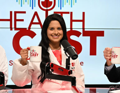
Description of Video Studio: News desk with Stony Brook Medicine logo on the front. A big screen is behind seated experts with the Healthcast logo (red uppercase lettering with a microphone at the top of the “L”). Music plays as the announcer introduces the episode.
Announcer
Welcome to Healthcast, where leaders and experts from Stony Brook Medicine come together to discuss a range of topics from leadership and strategic planning to patient care and the inner workings of a successful healthcare system.
Allison McLarty, MD
Hello everyone and welcome to Healthcast. My name is Dr. Allison McLarty and today we’re discussing heart valve disease and how it impacts well-being and survival.
As the severity of valve disease increases, it can cause a range of symptoms such as heart failure, like dyspnea, orthopnea, arrhythmia, or syncopy or chest pain. And ultimately, severe valve disease left untreated is fatal.
However, most types of valve disorders can be treated effectively, and increasingly, we’re able to use state-of-the-art surgery to repair patients’ native valves, which can then restore their life expectancy back to near normal.
In a few minutes, we will be speaking with some of our surgical experts to discuss innovative surgical options to treat aortic and mitral valve pathologies. But first, let’s speak with Dr. Smadar Kort, who is the Director of Non-Invasive Cardiovascular Imaging at Stony Brook Medicine.
Accurate diagnosis of valve disease is critically important, and imaging is invaluable in procedural planning and measuring results. Dr Kort.
Smadar Kort, MD
Thank you, Allison. My name is Dr. Smadar Kort and I’m, like you said, Professor of Medicine at the Renaissance School of Medicine. I’m the system director for non-invasive cardiovascular imaging and echocardiography, and I’m also a co-director of the valve center.
1:55 Heart Valve Disease Diagnosis and Advances in Cardiovascular Imaging
Allison McLarty, MD
Okay, so definitely an expert, and that’s very helpful for us today. So can you tell us how heart valve disease is diagnosed, and what key advances have been seen in cardiovascular imaging recently?
Smadar Kort, MD
So the key tool that we have for diagnosing valvular heart disease is still echocardiography. However, the echo that we do today is very different than the echo that I was doing as a cardiology fellow years ago.
So we now routinely use 3D and 4D for assessment of valve anatomy, also for more accurate assessment of cardiac volumes – left ventricular volume, left anterior volume. We utilize strain imaging as a more sensitive parameter for assessment of systolic function, and we have all sorts of formulas of calculations to really assess the degree of severity.
So when we talk about diagnosing valvular heart disease, obviously we need to assess the anatomy, we need to visualize the valve, we try to figure out the etiology of the valve pathology.
We then assess the hemodynamics so we can quantify the severity of that particular valve disease. We then look at the effect of that particular valve disease on the rest of the heart. So we really need to have a very accurate assessment of LV, size, LA size, assessment of the right side, peer pressures, as well as systolic function and diastolic function.
So this is how we essentially start with the transthoracic echocardiogram. Oftentimes we perform stress echo, so we put a patient or a person on the treadmill, and we get echo pictures before as well as after, and we compare them. And that’s extremely helpful for either patients who have exertional symptoms, we really try to see what happens with their valvular function as well as their LV function with exertion and it’s also extremely useful for patients who have discrepancy between resting images and their symptoms.
So if we have a patient that is very symptomatic, but their resting echocardiogram is not really demonstrating severe enough pathology to explain their symptoms, we exercise them and we see what happens to their valve disease, what happens to their peer pressures with exercise.
Likewise, if we have patients who have mildroid disease, and we want to see how they’re going to be doing under stress, so we we have seen many patients at Stony Brook with mitral stenosis, and the question is, are they going to be able to get pregnant, and how well are they going to be doing? Do we need to intervene before pregnancy?
We put those patients on a treadmill, and we can see what’s going to happen to their peer pressures, what is going to happen to the gradient supposed to valve, and that gives us an indication in terms of how well they’re going to be able to tolerate pregnancy or any other stressful event.
Allison McLarty, MD
That’s amazing. That definitely is a huge change from the way it was when I was training also. So a lot has happened in the last 10 to 20 years with echo.
So with such a comprehensive examination like that, how long would a patient expect to be on the echo table for your team to complete such a thorough study.
Smadar Kort, MD
So our lab is an accredited lab, so we really go by national guidelines, and we get pretty much between 45 minutes to an hour for an exam. And I can tell you that we typically do utilize that. For valvular patients, we do perform all the quantifications, all the calculations. Oftentimes, like I said, we do add 3D measurements as well. So it takes about an hour to perform a transthoracic echo on these patients. Transesophageal echo, which is a whole other modality, still utilizing echo, but with the additional probe, that’s extremely helpful, especially for assessment of the mitral valve.
So we performed the echo and for the mitral valve, really get the surgical view of the mitral valve. So we can really look at this valve the same way that you look at the valve in the OR and in this way, we can really look at the pictures together, and we can have very meaningful conversations about the anatomy, the pathology, the hemodynamics, and what needs to be done. So transesophageal echo is extremely helpful and we use quantification as well when we perform transesophageal echo.
7:11 Surgeon + Cardiologist Collaboration
Allison McLarty, MD
Yeah, I know that it has been extremely valuable for us as surgeons to sit with you as cardiologist and a cardiographer, so that, as you said, we’re looking at the images together, and we’re looking at the valve the way that we would see it in the operating room. And that way we can really pin down what the pathology is so that in the operating room, we can try to address that pathology specifically.
And also in the operating room for some of our procedures, having yourself and members of your team present during the procedure is also very critical, and this is especially so in some of our percutaneous therapies, like the mitral valve edge-to-edge repair.
So talk to us a little bit about what that kind of echo work is like in the operating room when we’re trying to get a valve percutaneously closed.
8:00 Pre-Procedural Planning and Personalized Treatment Plans
Smadar Kort, MD
So I would actually take a step back because before we even bring the patient to the hybrid OR, we have to make a decision about the timing of intervention. So we start by really assessing, is there a valvular disease, what it is, the severity of it, how it affects the rest of the heart, and then make a decision about the timing, because we do not want to intervene too early. Yet, we do not want to wait too long before there is damage which is irreversible.
So that’s a big part of what we really do and this is when we use all those other tools like strain imaging, getting a stress test done, so we can really look at the patient’s hemodynamics, their exercise tolerance. We put it all into that equation and have a discussion, typically among all members of the team, as well as the patient and their family members, so we can all make a decision together about the best timing.
So before we actually bring the patient, once we decide that the patient would benefit from intervention, we need to decide what kind of an intervention, and here at Stony Brook, we have all options available, and we’re going to hear more from the surgeons and the interventional cardiologists about the various options. But we do have both surgical as well as non-surgical options for the patients. And within each one of them, we do have multiple different options, so we try to make that decision before we bring the patient for the procedure.
And we utilize echo, we utilize CT. It’s extremely helpful to reconstruct the heart with the new device or the new valve in place, we can actually see what it’s going to look like. And based on that, we can recommend some additional procedures and additional devices that can be used to really make the procedure more feasible and safer for the patient. So then we can present the patient with all that information, and by the time the patient shows up in the OR or hybrid OR, the operator knows exactly what is the planned procedure, and has everything available at their disposal to be able to perform that procedure.
So now that we’re in the hybrid OR…
Allison McLarty, MD
And actually, yeah, before you actually get into the hybrid OR and you’re going to tell us what that’s like. I just want to emphasize what you said about the preprocedural planning and the exhaustive steps that are taken to make sure that the diagnosis is accurate, the extent of disease is accurate, and that we’re very clear on what pathology needs to be fixed.
And I think a very important part of what we offer here at Stony Brook is the power of the multidisciplinary team. Because, as you said, you know, we the team, which involves yourself and interventional cardiologists and surgeons and so on, you know, we meet regularly to review these patients, and it’s very much a matter of measuring three or four times and then cutting once to make sure that when we actually do our procedure, we do it precisely, and we do it right, and we do it just once.
Smadar Kort, MD
Yes. So I think it is really exciting that we have all these therapeutic options. We have the diagnostic options, but we also have lots of therapeutic options for our patients, but we do need to choose the best one, the optimal one for the specific patient, and it is really tailored to the individual patient.
And this is why all these team interactions are really, really critical.
11:59 Hybrid OR and Real-Time Imaging
So now we’re in the hybrid OR to perform a procedure. And in the OR, we’re the heads of the interventional echocardiographer, which is a field within cardiology that clearly did not exist when I was a fellow. But essentially the way to describe that niche is that I’m essentially the eyes and the GPS of the procedure.
Yeah. So I use 3D TE, sometimes with additional eyes, with the intracardiac echo as well, and in which we utilize those to guide the procedure. So I can visualize all the wires, the catheters.
I look at the valve again, assess the pathology and guide interventional in terms of where to place the device. And if it’s a valve that is being repaired or replaced, then where exactly the repair needs to take place.
You mentioned TAVR, you mentioned the matra clips. So where the clip is supposed to be placed. And we have the advantage of actually assessing the effect of, let’s say, the clip before we deploy it. So that’s when I get my images, I do my calculations and measurements and assess the amount of residual leak, if there are any gradients across the valve. Is the valve now becoming too tight? Or do we have the ability to use more than one clip?
So again, this is not something that we can, we kind of have an idea before we go in, but oftentimes this is the fine tuning that is being done based on real time imaging in the hybrid OR.
Allison McLarty, MD
Which is critically important because without the eyes, we can’t see what we’re doing, and the procedure isn’t getting done properly. So it’s really important. And then, once a valve has been successfully repaired or replaced, what kind of surveillance is necessary after surgery to make sure that patients continue to do well?
14:05 Post-Op Surveillance
Smadar Kort, MD
So the goal is always for the patient to outlive the device or the valve that we put in place. So the first thing that we need to do is establish a first baseline for the patient. So that’s the full echo that we do just prior to discharge from the hospital. And typically, the patients stay here overnight just to monitor them. So before they go home, they get a full echocardiogram. We assess not just the valve that was repaired or replaced, but we really look at everything, because affecting one valve or treating one valve can affect the rest of the heart.
So we reassess their LV function and the RV function, and we measure the peer pressures, and we assess for any residual leak, any stenosis, and this is now becoming the new baseline for that patient. And most of our patients stay within the system, so they come back to us for follow up echoes, and we look for progression.
We also need to keep in mind that with the percutaneous or transcatheter procedures, we typically address one valve at a time. We don’t really, it’s not like an open heart surgery that we fixed everything that is abnormal. We make a decision about what is really the most critical valve that needs to be addressed first, we address that with the hope that maybe if we’re going to address one valve, the other valves may improve in function.
So we do need to bring those patients back, and we look at the other valves and look for either progression or improvement in the function of those valves. And we also look for progression or deterioration of the new valve that we put in place, or the new device that we put in place. So follow up is extremely, extremely important.
Allison McLarty, MD
Is there anything else that you’d like to add before we wrap up?
16:06 Importance of Screening for People with Elevated Risk Factors
Smadar Kort, MD
So we typically talk about people that have valvular heart disease and how we address them and how we diagnose them. But I really want to emphasize that it is also very important to at least get an echocardiogram on people that are at risk for developing valvular heart disease.
And those would be either people who have family members who were diagnosed with valvular heart disease. The most common one would be a bicuspid valve. Also people that have non valvular heart disease, like cardiomyopathies, ischemic, non ischemic cardiomyopathies can have secondary effects on the valves.
Also, patients who have other systemic disorders like rheumatological disorders, those patients are at risk for developing viral heart disease. So it is really important, especially for primary care physicians, to really think about those people and refer them to us whenever they are at risk for developing valvular heart disease.
Allison McLarty, MD
What are the triggers in terms of age of patient, for example, that such a referral should take place?
Smadar Kort, MD
So for family members, if you have a patient at any age that a family member, first degree family member was diagnosed with either congenital heart disease or with a known valvular heart disease, like a bicuspid valve, you really want to gather all the first degree relatives and send them for one echocardiogram, and hopefully that will be sufficient to basically exclude the presence of any other congenital anomalies or the presence of bicuspid valve, and that will be the first and last echo for them. But if you do find it, you really want to then follow them up, hopefully you would diagnose them early enough, so they would have no symptoms, and maybe we can intervene before there is any deterioration or any symptom development.
Allison McLarty, MD
That’s a very valuable piece of information, and I think something that many people don’t consider the importance of screening for this where there are elevated risk factors. So thank you very much for that.
We’ll be right back with Drs. Price and Yammine to continue our conversation.
18:45 Introduction of Surgical Experts
Welcome back, everyone. Now that we understand how valve disease is diagnosed, let’s turn to how we fix it, and for this, we are going to speak with our surgical experts. We’re fortunate to have with us today Dr. Jonathan Price, who is the co-director of the Aortic Center here at Stony Brook, and Dr. Maroun Yammine, who is the director of adult congenital surgery.
So Dr. Price, let’s start with you. We heard from Dr. Kort earlier about how we diagnose valve disease. Can you speak a little bit about how we fix it surgically?
19:06 Treatments for Heart Valve Disease: Valve Repair Surgery v. Valve Replacement
Jonathan Price, MD
Sure. So I think once you have a valve problem diagnosed by your doctor, it’s important to know that there are two broad ways to address it. One is to repair the valve. One is to replace the valve. I think the most important thing to remember for the patient is that at the end of the day, whatever you have to have, it’s to make your valve work and to make your heart pump better and more efficiently. So both modalities are options, but it’s based on a discussion with you and your doctor about which one is best for you.
Allison McLarty, MD
Okay, that’s fair enough. So Dr. Yammine, tell us, why would a doctor prefer to recommend valve repair surgery rather than valve replacement? Are the two treatment options equivalent, or are there advantages for one over the other?
Maroun Yammine, MD
So for certain specific patients, there is an advantage of repairing the valve, or what we call preserving the native valve, compared to replacing it with either a biologic prosthesis or an animal prosthesis, or a mechanical prosthesis or metallic. If patients receive a metallic prosthesis, they have to be on blood thinners the rest of their life. For those who have a biologic prosthesis, will have a certain lifespan of that prosthesis, and might need reintervention down the line.
For certain patients with certain problems, we can preserve their native valve, and by preserving it, that valve could last much longer than an animal valve, and it doesn’t need blood thinners like a metallic or a mechanical valve would. So that’s where the bigger advantage of preserving the native valve or repairing it comes compared to replacement.
Allison McLarty, MD
Okay, so that’s very helpful. So then Dr. Price, tell us what are some of the diagnoses that would require valve replacement rather than even consideration for valve repair?
Jonathan Price, MD
So again, in general terms, the valve cannot be repaired once the tissue of the valve has been destroyed. And in general, for the aortic valve, that would be aortic stenosis, when the valve is heavily calcified and deposited with sclerotic tissue, and the valve no longer opens properly.
In those cases, repairing the valve is not possible. Similar for the mitral valve, when you have mitral valve stenosis, not every case, but in most cases of mitral valve stenosis, when the leaflets are heavily calcified and restricted and not moving properly, those leaflets are not repairable anymore.
Allison McLarty, MD
21:54 Valve Replacement: Mechanical v. Biological Prostheses
Dr. Yammine mentioned mechanical prostheses and biological prostheses. So can you make a recommendation on which one might be better for a patient? Like, how does a surgeon decide which prosthesis to offer a patient, and how should a patient think about it and decide which one to accept?
Jonathan Price, MD
That’s an excellent question, and it’s really one for the ages, because I think that is one of the most heavily debated topics when physicians speak to each other and when physicians speak to patients, and when patients speak to patients.
And the reason is because everyone will have an opinion, and there is some good data to guide our recommendations. But at the same time, a valve replacement and the type of valve you use really is dependent on the patient, their lifestyle, comorbidities and a lot of other factors.
So for instance, as Dr. Yammine mentioned earlier, a metallic valve or a mechanical prosthesis, may not be something someone is interested in because they have to be on blood thinners for the rest of their life. However, a mechanical valve has a longevity and a likelihood of staying patent and lasting and working much longer than any other type of prosthesis.
Allison McLarty, MD
Like, how much longer?
Jonathan Price, MD
Well, decades longer, potentially. So the question then comes for a very young patient who might need a valve replacement, is a mechanical valve a viable option? And the answer is yes, it is a viable option because it’s still a very good operation that gives very good results. Now, the counter side to that is that you do have to be on blood thinners, and not just any blood thinner. As of right now, the only blood thinner that’s FDA approved to be used with a mechanical valve is Warfarin. And as most people know, Warfarin is labor intensive, time consuming and energy consuming. It takes a lot of effort on the part of the patient and the physician to make sure that it’s being used properly and making sure that it’s being effectively used so the patient’s blood thinner levels are appropriate for the type of valve.
That’s one thing that you cannot skimp on. If you have a mechanical valve, you need to be on your Warfarin, and you can’t take it lightly. You have to be checking regularly.
24:11 Valve Repair Options
Allison McLarty, MD
So, Dr. Yammine, Dr. Price just said that for a young patient, a mechanical valve might be a good option because it might last for many, many, many decades, and therefore not necessarily need to be replaced. So for a young patient who has a valve abnormality and is going to need surgery, can you speak a little bit about the valve repair options that might be possible that might obviate the need for mechanical valve and therefore the need for lifelong anticoagulation for this young patient, for many, many, many years?
Maroun Yammine, MD
Absolutely. I think before going into valve repair, it’s very important to point out that, as Dr. Price mentioned, every patient would benefit from different surgeries. Some patients have more than one option, and we should discuss all options as some of and then, depending on each patient, their best option might be different. Some patients, where the valve is repairable, we can offer repair. Some patients, especially young ones who need the replacement, mechanical prostheses are a great option. Biologic prostheses have their limitations.
If a patient is found to be a candidate for valve preservation or valve repair, then it is possible for both aortic valve and mitral valve disease. It’s more commonly done for mitral valve because it’s a more common problem that we see, and it’s been there for a much longer time compared to the aortic valve repair. But aortic valve repair is also known, and it’s studied and has good long term outcomes. We have up to 10 years follow up for the aortic valve repair, and usually younger patients with aortic regurgitation would benefit from it. But there are certain strict criteria for repair, which makes some patients unfortunately not candidates for it, and then we’d have to look into other types of replacements.
26:12 Surgical Techniques to Achieve Repair or Replacement
Allison McLarty, MD
I see, okay, fair enough. Now, we’ve heard a little bit about valve replacement. What about the surgical techniques that one would use to achieve either replacement or repair? You know, what would a referring doctor be thinking about when they’re sending their patients to us to be treated?
Jonathan Price, MD
So I think again, it depends on the patient, it depends on what comorbidities they have, and it depends on what we’re trying to achieve with the valve replacement or repair. You know, we have many options. Open heart surgery is an option. Open heart surgery can be done with a sternotomy, a full incision. It can be done with a mini sternotomy, or basically a mini incision. It can be done through a thoracotomy, through the ribs.
So there are different ways to access the heart and to get a valve implanted. It doesn’t always have to be the traditional way that people most think about when they think of open heart surgery. And again, depending on what the patient needs, many patients would be, I would say, a candidate for different access points.
Allison McLarty, MD
Which is good because, as you know, the world is moving towards minimally invasive interventions. And obviously, if we can minimize the invasiveness of what we’re doing to our patients, we can hopefully accelerate recovery.
So Dr. Yammine, can you speak a little bit about some of the very, very minimally invasive options that are available, including things like transfemoral percutaneous options for the aortic valve, the mitral valve, and actually now the tricuspid valve?
27:41 Minimally-Invasive Options
Maroun Yammine, MD
So percutaneous options for different valves have gone through different generations and advancements and the latest form now, we know that the treatment for aortic valve disease that is the TAVR, which is percutaneous, that has come a long way, and it’s become one of the most common procedures we do with, along with our colleagues from intervention cardiology. That one is used very commonly to treat aortic valve stenosis, where the valve is not repairable. And it’s a procedure performed through the groin with catheters, and where the aortic valve is replaced.
For the mitral valve, there is a mitral clip, which is another procedure we perform, and the tricuspid valve as tricuspid clip, where both valves are not replaced, but rather repaired or reserved, just in a way to lower the amount of regurgitation or backwards leak that those valves are causing to help the patients feel better and recover from their disease.
Allison McLarty, MD
So you know, if, if I was a patient, I would want the most minimally invasive option. So, Dr. Price, tell me, why not offer all patients percutaneous options like the TAVR or the percutaneous valve, mitral valve and tricuspid valve repairs that Dr. Yammine just described. Like what might be exclusion criteria for not having this minimally invasive option. Because if you can avoid an incision and open heart surgery, then why not go for it again?
Jonathan Price, MD
An excellent question. Because, you know, that is the thing that people ask us the most. Why would I ever want surgery if I could just have a puncture hole in my groin? And the fact of the matter is that not every minimally invasive option is good for every patient.
Now you asked specifically what kind of criteria we look for, and it depends on the valves. So let’s start with the aortic valve. When you’re talking about an aortic valve replacement, let’s say open surgery, versus a TAVR, which is basically a stent with a valve implanted in it that is inserted through the groin. There are both options to replace a stenotic valve or the pathology of aortic stenosis. However, there are certain pathologies, or, you know, extents of aortic stenosis and calcification and valve morphology that makes the TAVR less likely to function properly.
So for instance, a very heavily calcified aortic valve, or when the calcium extends off the valve but down into the left ventricle, those are things that make the risk of injury to the heart and or a an improper seal of the valve, such that there is paravalvular regurgitation much more likely.
And if you’re talking about a young patient who is coming to you for a perfect outcome, to trade one pathology for another, and a young patient seems to be not imperfect.
Allison McLarty, MD
And define young, because, you know, like 60 is the new 50, if you will. What’s young these days?
Jonathan Price, MD
So again, it’s interesting because we do open heart surgery on people in their 80s. We also do TAVRs on people in their 50s, and it really depends on the patient and their risk profile for surgery, and whether or not we think that they will benefit from the surgery.
Now, in planning for this, you know, we have a lot more long term data with surgical valves than we do with TAVR valves. So, you know, the TAVR procedure has only been around for about 20 years now, and, you know, being performed here at this institution in the northeast and most of the hospitals in the area for, you know, a little over a decade.
So the fact of the matter is, the long term data really isn’t there to support, you know, do you have 10 or 20 years of longevity out of these valves? Now, we do know the surgically placed valves, especially the bioprosthetic valves, do not last forever. They also have a shelf life, but we do at least have the data to report that. With the TAVRs we really don’t. So for a young patient who is looking for a certain outcome, we would likely offer them surgery over a TAVR, because the longevity is not, not quite clear.
That being said, certain other types of valve pathologies, like people who have bicuspid valves. A bicuspid valve causes an abnormal geometric shape of the aortic root. And when you place a perfectly rounded aortic valve stent in an abnormally shaped root or an oblong root, there is a higher chance that you will have a paravalvular leak, which, again, may not be ideal for certain patients.
So those are things we all take into consideration when we discuss with the patient what would be better for them. Because obviously, no one wants surgery, but at the end of the day, that may actually be the thing that saves their life and is really the best thing for them.
32:49 Working with Problematic TAVR Valves
Allison McLarty, MD
And you know, Dr. Yammine, in the early days of the percutaneous valve therapy, especially TAVR, there was a lot of enthusiasm. There were low risk trials and a lot of low risk younger patients had TAVRs. And as you gentlemen know, we have seen a spike in patients with complications of their TAVRs coming now for open heart surgery. Can you speak a little bit about some of the complexity of trying to deal with a patient with a problematic TAVR valve who now needs surgery?
Maroun Yammine, MD
So yeah, having a previous manipulation to the valve, especially with having a new replaced valve in, would make the surgery for replacement or re-replacement more complicated. It’s always the case. Second time operation is always more complicated.
Now we have a lot more experience in doing second time operations following a previous surgical replacement. But as we’re gaining more exposure and experience with the trans catheter, we’re realizing that it does cause little more, maybe damage to the aorta around it to the aortic root, and sometimes we find ourselves having to repair the aorta around the TAVR valve before we implant the new aortic valve prosthesis surgically. So there’s no doubt it makes it more complicated, but it’s something that we’ve gained experience in, and we’re very comfortable offering to our patients if that’s the procedure they need.
34:38 Multidisciplinary Approach to Valve Repair at Stony Brook Medicine
Allison McLarty, MD
Alright, so we’re going to be winding up soon. Do you have any final thoughts or comments that you’d like to make about the spectrum of valve care that we can offer here at Stony Brook? We’ve heard a little bit about surgery and incisions and percutaneous options. I will just say that what we also have is an excellent multidisciplinary team that we can sit with to discuss complex patients. Anything else that you’d like to highlight about the services that we offer here.
Jonathan Price, MD
Absolutely. So I think that’s a great point to bring up, the multidisciplinary team, because at the end of the day, I think the overarching theme of this entire talk is that there are a lot of different options for valve intervention, whether it be a repair or replacement, and every patient deserves an in depth conversation to see what is right for them. And it’s important to know that that decision is not made unilaterally by the surgeon or a cardiologist or a referring Doctor.
This is really a conversation that happens with many, many physicians, who are each giving their unique expertise and insight to make sure that no stone is left unturned, and the patient is really being evaluated for every single option. And at the end of the day, when there are multiple options, and the patient has all of the data and all of the information available to them, they can choose what they think is best for them.
Allison McLarty, MD
Right, and I think being able to offer patients choice is important. Sometimes too much choice gets confusing. So I think we do need to be able to at least give them some structure within which to make decisions.
So on the heels of talking about multidisciplinary decision making and selection perhaps, Dr. Yammine, we could end with you just describing a little bit about one of the more complex decision making options for patients, which is the introduction of something called the Ross Procedure. Tell us a little bit about it and why a patient might be a candidate for this kind of valve surgery.
36:17 The Ross Procedure
So the ROS Procedure is a procedure that was initially performed and invented for congenital aortic valve disease, or it happened in neonates or kids with aortic valve disease, where they cannot fit in a prosthesis that is either the metallic or the biologic. So it’s a more complicated procedure where the pulmonary valve, or the one of the other valves in the heart is moved to the aortic position to replace the aortic valve. And in kids, it was shown to work exactly like an aortic valve, and it grew with these patients to adulthood.
So that had been applied to adults with aortic valve disease, especially those in the younger age group. So in fact, it’s actually considered in the American College of Cardiology and American Heart Association as the procedure that’s a possible option for patients who are younger than 50 to 55 years of age. It has the benefits of not having a metallic prosthesis or mechanical, so no anticoagulation. It certainly lasts much longer than an aortic valve that is bioprocess or biologic or animal based prosthesis. The only thing is that it’s a more complicated procedure, so it cannot be offered in all institutions, only institutions with good experience with it.
So we’re very proud to be able to offer this here at Stony Brook. And it’s definitely more complicated to decide on doing this procedure, and the technicality of the procedure is more complex than in any other aortic valve replacement, but it’s very well described and has been there for more than 30 years now.
But yeah, it adds to the complexity, and especially as Dr. Price was saying earlier, that the age group plays a major role in deciding which prosthesis or which choice to make. The guidelines do help us by saying that if you’re less than 65, you should have the surgery considered, even though tavern might still be an option, but as you move further younger than 65, as you get closer to the 50s, or even younger than 50s, that’s when Ross versus mechanical, possibly biologic, becomes more of a conversation to have.
39:10 Closing Remarks
Allison McLarty, MD
Okay, all right, that’s very helpful, and that’s all the time we have today. Thank you very much to all our panelists, and thank you to all our listeners and viewers. As we’ve heard Stony Brook Medicine’s Heart Institute offers the full range of treatment options for heart valve disease, and we prioritize repairing native valves where possible to confer the greatest long term benefit to our patients.
Our outstanding multidisciplinary team will care for you or your loved one with valve disease from diagnosis to discharge and provide state of the art treatment to get you back on your feet in a timely fashion. For evaluation by one of our team, please call (631) 444-1820,
and if you found this information helpful or informative, please like and subscribe for more content just like this.
Announcer
Stony Brook Medicine is Long Island’s premier academic medical center. We transform lives through scientific discovery, education and care, and we bring together innovative research, advanced education and extraordinary healthcare expertise to set the standard for how healthy communities thrive. For more information, visit stonybrookmedicine.edu or follow us on social media.