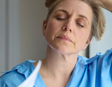

According to the American Cancer Society, breast cancer is the most common cancer diagnosed among women in the United States. It is also the second-leading cause of death from cancer among women. Regular screenings are critical for early detection and successful treatment. In this episode, experts from Stony Brook Medicine dive into breast health essentials, including risk factors, screenings (including after a breast cancer diagnosis), breast density and more.
The Experts
What You’ll Learn in This Episode
- 00:00 Opening and Introductions
- 2:04 Importance of regular breast exams
- 3:36 Risk factors and family history
- 6:38 Screening mammogram recommendations
- 7:22 2D v. 3D Mammograms
- 8:05 What to expect with a 3D mammogram
- 9:00 Mammograms and radiation exposure
- 9:50 False positives
- 11:13 Breast density
- 14:32 Screenings with implants
- 16:22 Screening MRI
- 18:10 Screenings after breast cancer
- 21:52 Closing Remarks
Full Podcast Transcript
00:00 Opening and Introductions
Description of Video Studio: News desk with Stony Brook Medicine logo on the front. A big screen is behind seated experts with the HEALTH Yeah! logo (red uppercase lettering with a microphone at the top of the “L”). Music plays as the announcer introduces the episode.
Welcome to HEALTH Yeah!, where experts from Stony Brook Medicine come together to discuss topics ranging from the complex inner workings of an infectious disease to tips and tricks for staying safe and healthy all year long.
Tara Huston, MD, FACS
Hello and welcome to HEALTH Yeah!. My name is Dr Tara Huston and today we’re going to talk about all things breast health, from risk factors and screenings to mastectomy post reconstruction and more. Whether you’re preparing for your first exam or navigating life after mastectomy, this episode is packed with expert insights and tips to support you in your breast health journey. Let’s get started.
I have with me today Doctors Anastasia Bakoulis and Cindy Lee. I’m going to ask each of you to take a moment to introduce yourselves to our listeners and viewers. Dr. Bakoulis, let’s start with you.
Anastasia Bakoulis, DO
Hi, Dr Huston, thanks for having me. Hi, Dr. Lee. I’m Anastasia Bakoulis. I’m a breast surgeon here at Stony Brook. I’ve been here for close to eight years now, and I specialize in benign and malignant diseases of the breast.
Cindy Lee, MD, FSBI, FACMQ
Hi, thank you for having me. My name is Dr. Cindy Lee. I’m the Chief of Breast Imaging here at Stony Brook. I am the person who’s responsible for reading your yearly mammogram, taking care of you when you have an abnormal finding, and also performing the biopsy if you need it. So thank you for having me.
Tara Huston, MD, FACS
Thank you both. For many years, I’ve had the privilege of treating women in our community with breast cancer. These are fellow moms, colleagues and friends, as well as their mothers, daughters and sisters. Many of these women were fortunate to have had access to care. For most screening mammograms afforded early detection. This was not the case for all.
Breast cancer affects one in eight of us. I am honored to have become a part of their journey and driven to improve the access to and quality of breast care in our community. The purpose of this podcast is to educate and empower.
2:04 Importance of Regular Breast Exams
Let’s start with Dr. Bakoulis. Tell us about the importance of regular breast exams, self exams, as well as clinical exams by your physician.
Anastasia Bakoulis, DO
Sure. Self awareness, or the awareness of one’s own breast tissue, the skin, the nature of one’s nipples, is really important in knowing or recognizing any differences that may come up in the future. That’s what we mean by self breast exams.
The best time to perform self breast exams are in the time right after one gets their period, so after you start to bleed. This is usually because breast tissue is less tender at that point in time and also least dynamic, so the tissue texture changes that one could potentially appreciate in the days prior to the period can be very different.
You need not perform a self breast exam more than once a month. Some people feel more comfortable with twice a month. But really, once a month is enough.
So there’s no right way to do a self breast exam, but being consistent about how you do it is the best way. You can do it upright. You can do it with your arm over your head. You can do it in the shower, as your skin is lathered up with soap. Sometimes it’s easier that way. You can do it laying in bed.
3:36 Risk Factors and Family History
Tara Huston, MD, FACS
Dr. Bakoulis, regarding risk factors and family history, who should be the most vigilant?
Anastasia Bakoulis, DO
It’s a great question, Dr. Huston. Folks who have significant family history of cancer, and you know, we always ask about breast and ovarian but different types of cancer matter too. So individuals who have a significant family history, whether it be in their parents, first cousins, aunts and uncles, those are the folks who should be a little more wary of their history and their risk factors.
Individuals who’ve had a series of biopsies in the past, that tends to increase a person’s at least calculated risk of breast cancer. And then, there are other factors, like prior history of chest radiation as a young person. These are prominent risk factors to take into consideration.
Cindy Lee, MD, FSBI, FACMQ
I totally agree with Dr. Bakoulis, so breast cancer risk is additive, right? So we’re adding all of your risk factors from the family history, your personal history, and also your prior treatment history, so everything gets added together to calculate your personal lifetime risk of breast cancer.
Anastasia Bakoulis, DO
That’s right, Dr. Lee. On a daily basis in our practice, we use these nifty calculators when we meet folks who do have some notable risk factors. The most commonly used calculator is the Tyrer-Cuzick calculator. This is readily found online; you can easily just pull it up on a Google search. But this is a very useful tool to help identify people who are at higher risk for development of breast cancer, and there are things that we can do to them if that calculation is high.
6:38 Screening Mammogram Recommendations
Tara Huston, MD, FACS
So knowing that our greatest risk is being female, and then followed by all the risk factors that both of you just discussed, at what age would you like to see a woman come to you for her first screening mammogram?
Cindy Lee, MD, FSBI, FACMQ
So per the recommendation of the American College of Radiology and the American Cancer Society, we recommend annual screening mammograms starting at 40. But if you have a higher risk factor, for example, family history, personal history of breast cancer or even genetic mutation, you have to start earlier.
So your screening mammogram would then start yearly at 30, in addition to having supplemental imaging exams such as ultrasound and breast MRI.
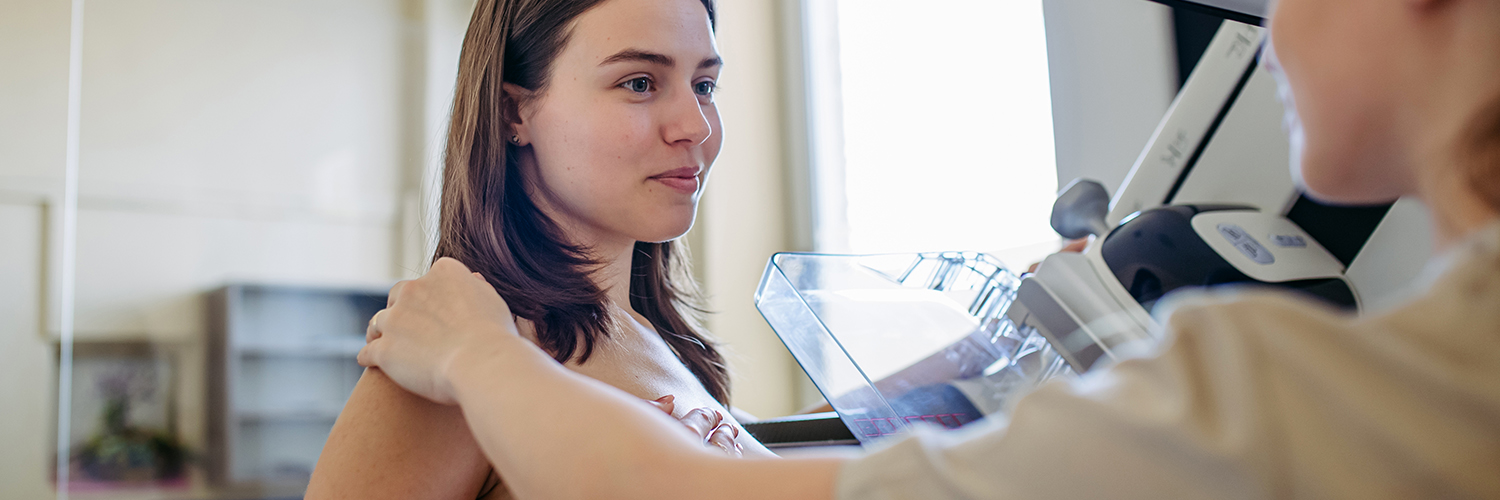
7:22 2D v. 3D Mammograms
Tara Huston, MD, FACS
Well, let’s start with mammograms. Something that a lot of women want to know is the difference between a 2D and A 3D mammogram. We see this advertised.
Cindy Lee, MD, FSBI, FACMQ
So there’s a huge difference in terms of technology and how much better the 3D machine is. So think of it this way, 2D only gives you one picture, one two-dimensional, and that’s it, where 3D gives you multiple pictures, like a video to your breast on the top to the bottom, from the right to the left, and side to side. So it really gives you so much more accurate information and allows us to find earlier cancer at a smaller size and earlier stage.
So it really makes a difference to make sure you’re getting your 3D mammogram this year.
8:05 What to Expect with a 3D Mammogram
Tara Huston, MD, FACS
So tell our viewers what to expect when they make their appointment for a 3D mammogram and then they come into your office.
Cindy Lee, MD, FSBI, FACMQ
Okay. Number one, the patient usually is worried about pain and discomfort. It is not comfortable. So if the mammogram is done right, you should see a little bit of bruising. I typically do when I’m done with my yearly mammogram, and that’s okay, because of that two minute discomfort, you’re saving yourself years and years of early diagnosis. You have better quality of life, so it’s definitely worth it.
It’s just like a pap smear. No one wants it. No one loves it. But this is not invasive. When you come for your appointment, what we do is you’ll be changing into a gown, and we will position you correctly on the mammogram machine, and then you’ll have some pressure applied to your breast while we take a picture, and that’s it.
9:00 Mammograms and Radiation Exposure
Tara Huston, MD, FACS
Some other fears that women have shared are concerns for radiation exposure. And some women say, well, if I have a mammogram, is that going to give me breast cancer?
Cindy Lee, MD, FSBI, FACMQ
That’s my favorite question. So that’s very commonly asked by patients. So the radiation equivalent of a mammogram is like a chest X ray, right? And it’s also equivalent of someone who takes a flight from New York to California, and that background cosmic radiation you’re getting is the equivalent of a mammogram. And there are people who do that professionally, ongoing throughout their life, and they do not have increased cancer risk.
So my point is, it does not increase your cancer risk. It is a known minimal radiation, and it’s definitely going to improve the outcome.
9:50 False Positives
Tara Huston, MD, FACS
Dr. Lee, tell me about false positives. How often does that happen and what does it entail?
Cindy Lee, MD, FSBI, FACMQ
So false positives happen when we bring you back for additional imaging but do not find cancer. So that happens from time to time, because maybe you don’t have a prior mammogram, this is the very first time we’re meeting you. And sometimes it happens because you have dense breasts. Sometimes it happens because you made a new cyst, or truly have suspicious finding in the breast.
It definitely happens. I had a false positive once, but I am happy to be there to get the additional imaging for that peace of mind, so I could be sure of all of my imaging on that second visit, I could get to talk to the radiologist, and they can assure me that everything is good.
Tara Huston, MD, FACS
You mentioned more false positives when you’re having your first mammogram at a new facility. So is it better if women find the facility that they’re comfortable with and come back to the same facility and the same radiologist year after year? Does that help you do their read?
Cindy Lee, MD, FSBI, FACMQ
So what helps the most is the access to prior mammograms. Let’s say you went somewhere else for your first time, and then you decide to come to Stony Brook to have your mammogram second. I strongly advise you to get that outside picture and then bring it to us. That way you could be interpreted the same way as somebody who has been done here to minimize your false positives.
11:13 Breast Density
Tara Huston, MD, FACS
Dr. Bakoulis, let’s talk about breast density. What does that mean from an anatomic perspective?
Anastasia Bakoulis, DO
Sure, breast density is a very normal, physiologic thing. The density is not something that you feel. Density is something that you can see, mostly on a mammogram.
Breast density is a reflection of how much functional breast gland there is in the actual breast. So a breast is made up basically of a couple of layers. You have the skin on the outside, a layer of fat that is very normal right underneath that, then you have glandular tissue, which is a combination of ductal tissue and the milk producing factory of the breast, and then you have stroma, so that you have the scaffolding part of the breast. Breast density is a reflection of that functional part of the breast tissue.
As we get older, and as our body sees less and less estrogen as a result of menopause, that breast tissue does eventually go away, and as a result, our breast tissue becomes less and less dense, which makes a mammogram even better.
So density is not something that you feel necessarily, and it’s a very normal thing in a person who is getting a very regular period. It’s even normal in people who haven’t had a period in a couple of years. But it’s the actual functional breast tissue.
Tara Huston, MD, FACS
And Dr. Lee, how did dense breasts affect the imaging modality that you choose and how you read those?
Cindy Lee, MD, FSBI, FACMQ
So I wanted to also put this in context of how many people have dense breasts in the US. So as radiologists, we categorize your breast tissue into four different kinds of density. We call it A, B, C and D.
If you have C category for heterogeneously dense breasts and also D, extremely dense breasts, then we consider you having dense breasts. So in the US population, typically, about 40 percent of women will have a category C, about 10 percent will have category D. So all together, it means half of US women will have dense breasts. So this affects a lot of women, a lot of the population in the US and also throughout the world. This is a really important topic.
So in terms of what can you do about dense tissue? Number one, this is normal, just like Dr. Bakoulis said, it’s physiologic. This is part of you. There’s nothing wrong with you having dense breasts, and number two, how do you take care of yourself if you do have dense breasts?
What we typically recommend is to do supplemental screening. So to begin with, that is ultrasound. Breast ultrasound is really great at imaging the dense tissue. For mammograms, sensitivity decreases as breast tissue gets denser, and the ability for me to see your cancer decreases on mammogram.
So typically, the first step we will recommend for an additional test is breast ultrasound. And if you do have other risk factors, and your lifetime risk is over 20 percent, then breast MRI will be indicated as well.
14:32 Screenings with Implants
Tara Huston, MD, FACS
So as a plastic surgeon, I often get asked about implants and if your mammographic screening is different when you do have implants. So can you talk a little bit about how you image the breast and the implant in a woman who either has implants for cosmetic augmentation or in a woman who has implants for reconstruction after mastectomy?
Cindy Lee, MD, FSBI, FACMQ
Absolutely, great question. So implants require special care. We do specialized views to displace we call them implant displacement view to push the implant out of the way of the mammogram machine. And they are a little bit tricky, so you do get more pictures taken so that we can see the breast tissue that isn’t a part of the implant.
Tara Huston, MD, FACS
As someone who screens implants for many years after placing them in, I know our guidelines changed in coordination with the American College of Radiology about five, six years ago, such that when implants are placed, be it for cosmetic augmentation or after we reconstruct following mastectomy, that we send our patients to you for an ultrasound at five years.
And if you’re happy with the way the ultrasound looks, and you tell us the implant is intact, we stop there, and then we send them back to you in two to three years for another ultrasound. But if anything abnormal is found on that ultrasound, then we ask that you do an MRI of the implant.
Cindy Lee, MD, FSBI, FACMQ
So especially for silicone breast implant, which the majority of breast implants are made of, they are difficult to see on the ultrasound, the sound wave actually does not penetrate through the capsule of your silicone breast implant. So what’s the best exam? The best imaging for us for checking your silicone breast implant is deep breast MRI.
16:22 Screening MRI
Tara Huston, MD, FACS
So let’s talk about MRI for a minute since we’re on that topic, and I know that Dr. Bakoulis likes to screen her high risk patients with MRIs as well. So I want to hear from both of you when you order and when you would recommend that a woman with her native breast, not implants anymore, have a screening MRI.
Anastasia Bakoulis, DO
Sure. So this is where genetic testing results are taken into consideration, Tyrer-Cuzick or other lifetime risk calculators, and their results come into play.
So you know, outside of a known genetic mutation that’s related to a breast cancer, if a patient is identified to have a greater than 20 percent lifetime risk of breast cancer, this is where breast MRIs come into play big time for us.
So in addition to conventional mammograms, plus or minus ultrasounds, depending on breast density, we alternate with breast MRIs, and we plan for this every six months. So if a person usually gets her mammogram and ultrasound in January, we would plan for a breast MRI for screening purposes in July. And so this way, every six months, something different is looking at the breast tissue. And it’s a really wonderful way, a very complete way, in my opinion, to analyze breast tissue.
Cindy Lee, MD, FSBI, FACMQ
And I want to add that breast MRI is actually one of the most sensitive tools we have in finding breast cancer. It is good at finding anything that could be cancerous, even things that are not visible on mammogram, ultrasound.
18:10 Screenings After Breast Cancer
Tara Huston, MD, FACS
Absolutely. Moving forward to treatment after breast cancer. When a woman has breast conservation therapy, when lumpectomy and radiation spares her breast, how often do you want that breast image?
And now she’s at a slightly higher risk in the contralateral breast. How often do you want the other breast image as well?
Anastasia Bakoulis, DO
Dr. Huston, that’s a really good question. And you know, I do look to my radiologist for a lot of guidance. I think most breast surgeons will image the affected breast after a lumpectomy and radiation in about six months after diagnosis, and then subsequently you resume your annual bilateral or both side screening.
MRIs are a little more controversial. I definitely include MRIs as part of my screening, if we find additional findings on the primary breast MRI at time of diagnosis. I think that would be very, very important, right?
Obviously, for an individual with a genetic mutation who pursued breast conservation therapy, an MRI is very important for those individuals. But I think you know an MRI annually after breast cancer diagnosis is not an absolute must, but certainly a conversation that you know doctor and patient should have on a regular basis.
Cindy Lee, MD, FSBI, FACMQ
And I think it really matters when the patient was diagnosed with breast cancer, and also, are there any other risk factors, such as your first degree family relative with breast cancer? How old were they when they were diagnosed? So all of that comes together and gets added together to calculate what you mentioned before is the 20 percent cutoff.
So if you’re clearly above the 20 percent risk for lifetime risk of breast cancer, then absolutely breast MRI is for you. But if you’re not above the 20 percent threshold, we don’t recommend it because it comes with rates of false positives. We don’t want to do any unnecessary imaging or biopsy if we can avoid it. So MRI is not for everyone.
Anastasia Bakoulis, DO
I can’t agree with you more. Dr. Lee.
Tara Huston, MD, FACS
Okay, I’m gonna complicate the screening question a little bit more. So we talked about how you’d follow up a patient who has implants, or implants even after mastectomy. What about a woman who has a latissimus flap, a TRAM flap, a deep flap after mastectomy, so has her own tissue and her newly reconstructed breast, but it’s not breast tissue. What type of screening would you recommend for that patient?
Cindy Lee, MD, FSBI, FACMQ
So for a patient who had a total mastectomy with no longer any residual breast tissue, then we do not recommend mammograms. Mammograms are really designed for imaging breast tissue, not flaps, not reconstruction breast tissue. So in those cases, if these patients are asymptomatic, ultrasound could be of use, and also breast MRI if they still have the other breasts.
Tara Huston, MD, FACS
Would you recommend breast MRI on the breast that’s had mastectomy and reconstruction, or just on the native breast?
Cindy Lee, MD, FSBI, FACMQ
Good question. Breast MRI is done bilaterally, meaning, whenever you go for MRI, we look at both, doesn’t matter what.
Tara Huston, MD, FACS
And Dr. Bakoulis, after we’ve reconstructed someone, how frequently should that woman be undergoing clinical breast exams by her breast surgeon, or plastic surgeon or her gynecologist?
Anastasia Bakoulis, DO
Sure. In general, we do recommend an annual exam, at least for the first five years, especially. And again, promote breast awareness, even though there isn’t really a native breast there anymore, but just a learning of what your new reconstructed tissues feel like now.
Cindy Lee, MD, FSBI, FACMQ
And then, if you were to have any new symptoms or a new palpable lump, you know, definitely come back. We can do an ultrasound. It does not have radiation; you do not need contrast, but it’s a great way to evaluate any concern you have.
Anastasia Bakoulis, DO
Absolutely, ultrasound, we can even do MRIs, like you mentioned earlier, there’s always a way to visualize things and investigate things, even after a mastectomy.
21:52 Closing Remarks
Tara Huston, MD, FACS
This has been a tremendous amount of information. If you had to leave our viewers with one key message or request. What would you say?
Anastasia Bakoulis, DO
You know, I think “know thyself” probably is the number one recommendation I’d make. I think it’s important. Women are moms, they’re caretakers, they’re busy people, and they’re always taking care of someone else other than themselves. So recognizing what is your normal is not the most natural thing sometimes. So know what your normal feels like. Know what your normal looks like.
And of course, if there’s any questions or if you have concerns, only you know you best, so reach out to your gynecologist, reach out to your doctor, reach out to your breast surgeon, right? And you know, if you’ve noticed any changes, it’s important to come forward with these concerns immediately.
Cindy Lee, MD, FSBI, FACMQ
I like that message. So my take home message for you all tuning in today is to make sure you did your screening this year. If not, we have a lot of different locations that’s convenient throughout different communities: Riverhead, Commack, Stony Brook, the hospital, Southampton, West Hampton. We have numerous locations designed to give you the most access and best equipment possible to help you get your screening done.
Tara Huston, MD, FACS
And my tip relates black plastic surgery again, and it’s “take care of your implants.” Implants need attention. They don’t always need to be changed. You don’t always need another operation right away, but it’s important to get your screenings. And if something feels abnormal to you, like you mentioned on your native breast, on your augmented breast, on your reconstructed breast, call one of us, and we’re here to figure it out and to help you.
So that’s all the time we have for today. Thank you so much to our panelists, and thank you to all of you, our listeners and viewers. Remember, screening is your best first step. For more resources, including information on screenings, you can call the number on your screen, and if you found this information interesting or helpful, please like and subscribe for more content, like this.
Announcer
Stony Brook Medicine is Long Island’s premier academic medical center. We transform lives through scientific discovery, education and care, and we bring together innovative research, advanced education and extraordinary healthcare expertise to set the standard for how healthy communities thrive. For more information. Visit stonybrookmedicine.edu or follow us on social media.
*DISCLAIMER: The information provided in this podcast is for educational and informational purposes only and is not intended as a substitute for professional medical advice, diagnosis or treatment. If you think you may have a medical emergency, call your doctor or emergency services immediately.